Anatomy Of The Eye
In the anatomy of the eye , three layers are distinguished: external, middle and internal. Likewise, the eyeball, which is what we popularly know as the eye, has several components inside it and also protective structures.
The outer layer of the eyeball is made up of the cornea and the sclera. The cornea is the outermost area of the eye’s anatomy. It is a transparent area, much like the glass on a wristwatch.
The cornea is made up of avascular tissue, that is, without blood supply. It is a highly specialized tissue that allows light to be refracted and transmitted to the retina, at the back of the eye.
The sclera, on the other hand, is an outer covering of a fibrous type that has a high content of collagen. It has a dense, whitish conformation. In the front, it continues with the cornea and in the back with the optic nerve. The eyeball moves thanks to the muscles that are attached to the sclera.
The anatomy of the eye in the middle layer
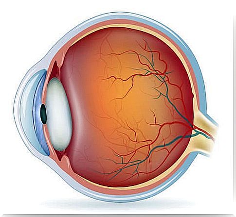
The middle layer of the eye is called the uvea and is made up of the choroid, the iris, and the ciliary body. Each of these components of the anatomy of the eye has a different conformation.
- Choroid: it is located between the sclera and the retina, nourishing the outermost areas of it. The choroid contains a pigment whose function is to absorb excess light and prevent blurred vision.
- Ciliary body: it is a structure of the anatomy of the eye that connects the choroid with the iris. It has a triangular shape and is the area in charge of producing the exit of the aqueous humor, through the Schlemm’s canal. It also causes the lens to change shape.
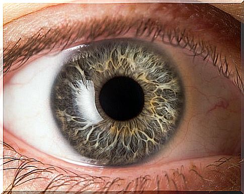
- Iris: this structure protects the eye from excessive light entry. It is located between the anterior chamber and the lens. In the center it has a circular opening called the pupil, which dilates or contracts. It can be of different colors, depending on the heritage.
The inner layer or retina

The inner layer of the wall of the eyeball is called the retina. In it are the cones and rods, which are the photoreceptors of the eye. This means that they allow us to perceive light and colors. In total, there are between six and seven million cones on the retina.
The cones allow to capture the colors during the day. In addition, there are about 120 million rods that make black and white vision possible in the dark. The photoreceptor signals are sent to the brain through the optic pathways found in the optic nerve.
The most important area is located in the center of the retina and is known as the macula or macula lutea . This area is responsible for giving color and sharpness to the images captured by the eye. It is the region of greatest visual acuity.
The inside of the eyeball
In the inner area of the eyeball are the following components:
- Crystalline: takes care of the sharpness and the focusing of light rays, along with the cornea. It is made up of a transparent fabric and surrounded by an elastic capsule.
- Anterior and posterior chamber : The anterior chamber is between the cornea and the iris. The posterior, between the iris, the lens and the ciliary body.
- Vitreous humor: it is a gelatinous tissue that occupies two-thirds of the eyeball. The vitreous humor is transparent and is 98% made up of water.
- Iridocorneal angle: it is formed by the cornea and the iris and allows the aqueous humor to drain.
Protective structures and optic nerve
The protective structures in the anatomy of the eye are:
- Bony orbits: they are concavities formed by seven bones adjacent to the skull. They have two openings, one at the bottom and the other at the back; nerves and veins pass through them. The eyeball occupies a fifth of the orbits and the remaining area has connective and fatty tissues, nerves, muscles and vessels.
- Eyelids: the eyelids are two mobile folds of skin, which protect the eye from external elements or strong glare. On the edge they have sebaceous glands.
- Lacrimal apparatus: The lacrimal gland is located under the outer part of the upper eyelid. It joins the nasal cavity through the lacrimal sac and the naso-lacrimal duct.
- Conjunctiva: it is a transparent and mucous membrane that covers the eyeball and protects it against germs and foreign bodies.
The optic nerve, for its part, is a cable that is formed from the confluence of the nerve fibers of the retina. These fibers number more than a million. It transmits visual impulses to the brain and connects the eye with the visual area of the brain.